Imaging in Fibroadenoma What Mammogram & USG Show
Breast swelling, heaviness, or lumpiness can be worrying for many women. One common benign (non-cancerous) cause of these symptoms is Fibroadenoma — a condition related to hormonal influence and fluid retention within breast tissue. While the symptoms can feel alarming, proper clinical evaluation and imaging help clearly identify the nature of the condition and rule out serious disease.
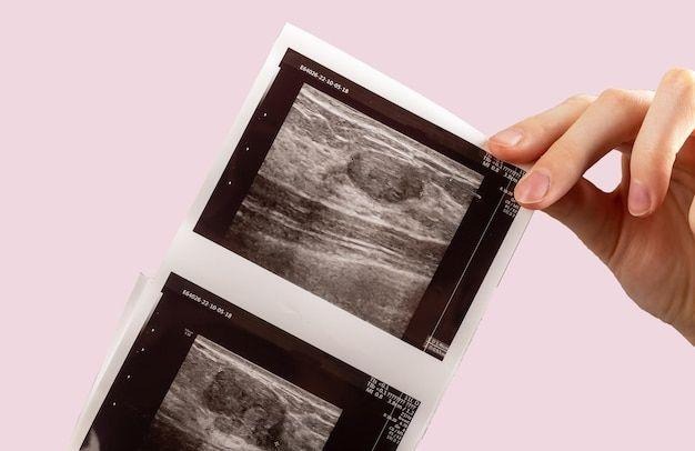
In this blog, let’s understand how mammography and ultrasound (USG) help doctors diagnose Fibroadenoma and what you can expect during imaging evaluation.
What is Fibroadenoma?
Fibroadenoma refers to hormone-related breast tissue swelling caused by fluid retention and fibrous tissue changes. It often presents with:
It is commonly seen in:
Because symptoms may mimic other breast conditions, imaging plays a key role in confirming the diagnosis.
Why Imaging is Important in Fibroadenoma
Clinical examination alone cannot always differentiate Fibroadenoma from:
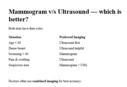
That is why doctors recommend:
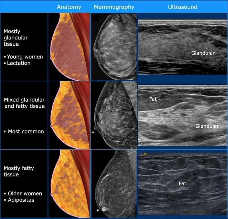
What Mammogram Shows in Fibroadenoma
A mammogram is an X-ray based imaging technique used mainly in women above 40 years or earlier if clinically required.
In Fibroadenoma mammogram may show:
✅ Increased Breast Density
✅ Symmetrical Changes
✅ No Spiculated Lesions
✅ Diffuse Pattern Instead of Focal Lump
However, mammography alone may not fully differentiate fluid from solid tissue — that’s where ultrasound helps.
What Breast Ultrasound (USG) Shows in Fibroadenoma
Ultrasound is extremely useful, especially in:
USG findings in Fibroadenoma commonly include:
✅ Increased Echogenic Fibroglandular Tissue
✅ Interstitial Fluid Changes
✅ No Solid Irregular Mass
✅ Possible Small Simple Cysts
✅ Normal Vascular Pattern (on Doppler)
When Additional Tests May Be Needed
Although Fibroadenoma is benign, further tests may be advised if:
Possible next steps:
Can Fibroadenoma Be Treated?
Yes — management is usually conservative:
Most cases improve with time and proper guidance.
When to See a Specialist
Do not ignore breast symptoms if you notice:
Early imaging provides reassurance and safety.
🏥 Advanced Breast Imaging at Bankers Vascular Hospital
At Bankers Vascular Hospital, we provide comprehensive breast evaluation with:
Our team focuses on accurate diagnosis, minimal anxiety, and evidence-based care — so benign conditions like Fibroadenoma are correctly identified and unnecessary procedures are avoided.


Comments
Post a Comment